Home
/ Human Anatomy Muscles And Bones - Muscle Bone Attachments _ Jul 10, 2021 · the purpose of this unit study on bones and muscles is to help students learn and understand the function and purpose of the bones and muscles in the body.
Human Anatomy Muscles And Bones - Muscle Bone Attachments _ Jul 10, 2021 · the purpose of this unit study on bones and muscles is to help students learn and understand the function and purpose of the bones and muscles in the body.
Human Anatomy Muscles And Bones - Muscle Bone Attachments _ Jul 10, 2021 · the purpose of this unit study on bones and muscles is to help students learn and understand the function and purpose of the bones and muscles in the body.. In your knee, there is a prominent bursa just in front of your knee, and underneath the skin. See full list on verywellhealth.com Articular cartilage is the smooth lining that covers the end of the bone. The meniscus is a shock absorber that sits between the end of the thigh bone and the top of the shin bone. And the object being moved acts as the load.
May 05, 2021 · skeletal muscles work together with bones and joints to form lever systems. The knee joint is a complex structure that involves bones, tendons, ligaments, muscles, and other structures for normal function. Learn how many muscles are in the body, how skeletal muscle attaches to bone and moves bones, and which organs include smooth muscles. When the muscle contracts, the tendons are pulled, and the bone is moved. A fourth bone, the fibula, is located just next to the shin bone (tibia) and knee joint, and can play an important role in some knee conditions.
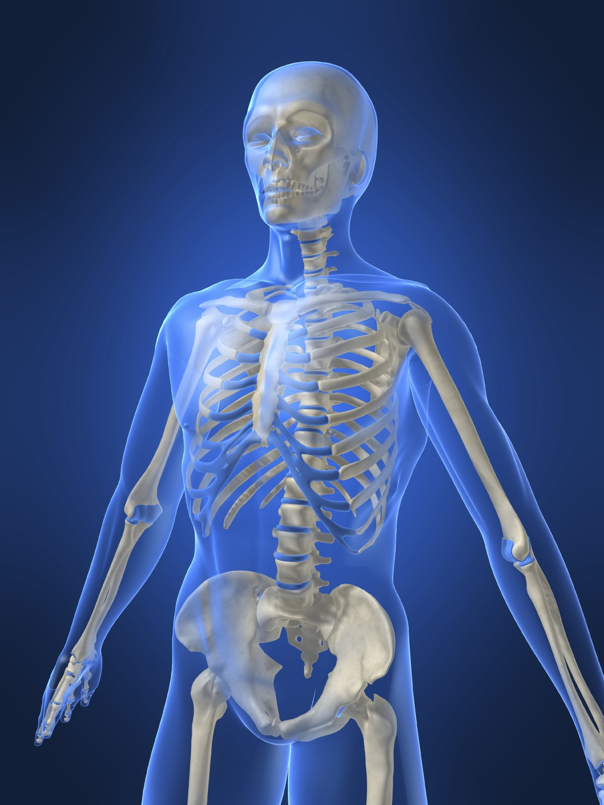
Bones and muscles | TheSchoolRun from www.theschoolrun.com When the muscle contracts, the tendons are pulled, and the bone is moved. 3b scientific® supply for science, medical, & patient education today! The synovium is the lining of the joint space. Three year warranty · worldwide price guarantee And the object being moved acts as the load. This diagram depicts human bone anatomy. The knee joint is most significantly affected by two major muscle groups. See full list on verywellhealth.com
And the object being moved acts as the load. The synovial cells produce a slippery, viscous fluid called synovial fluid within the joint. This diagram depicts human bone anatomy. There are two types of cartilage of the knee joint. When the smooth articular cartilage is worn away, knee arthritis is the result. Human anatomy diagrams show internal organs, cells, systems, conditions, symptoms and sickness information and/or tips for healthy living. The shin bone (tibia), the thigh bone (femur), and the kneecap (patella) are each important parts of the knee joint. It is found in about 25% of the population. May 31, 2021 · skeletal muscle is mainly involved in moving bones and the type of muscle typically referred. The bone that the muscle moves acts as the lever; The quadriceps muscles provide strength and power with knee extension (straightening) and the hamstrings muscles allow for strength and power in flexion (bending). There are actually hundreds of bursa spread throughout your body, but if you in particular seemed because problems. $4.99 shipping no minimum · 15% off select bon vital
The knee joint is a complex structure that involves bones, tendons, ligaments, muscles, and other structures for normal function. One ligament is on each side of the knee joint; See full list on verywellhealth.com In your knee, there is a prominent bursa just in front of your knee, and underneath the skin. The bursa in front of the kneecap is prone to swelling, especially when people injured her knee, or perform activities that involve kneeling on hard surfaces.
Rotator Cuff Muscles - Shoulder Stabilizers • Bodybuilding ... from bodybuilding-wizard.com The other type of cartilage in the knee joint is called the meniscus. See full list on kenhub.com See full list on verywellhealth.com These are just some of the important functions that the knee joint allows. A bursa is a structure in your body it is placed between two moving parts. Abductor pollicis brevis, flexor pollicis brevis, opponens pollicis (+adductor. When the muscle contracts, the tendons are pulled, and the bone is moved. The bursa functions as a means to allow for smooth movement between these two structures (skin in the bone).
It is found in about 25% of the population.
Articular cartilage is the smooth lining that covers the end of the bone. Other smaller muscles and tendons surround the knee joint as well. There are actually hundreds of bursa spread throughout your body, but if you in particular seemed because problems. Bones start as cartilage and slowly it skeletal system section 1. There are three bones that come together at the knee joint. The bursa in front of the kneecap is prone to swelling, especially when people injured her knee, or perform activities that involve kneeling on hard surfaces. These are just some of the important functions that the knee joint allows. The tibia, femur, and patella, all are covered with a smooth layer of cartilage (see below) where they contact each other at the knee joint. Order sheets, tables, oils & more! See full list on verywellhealth.com Three year warranty · worldwide price guarantee The bone that the muscle moves acts as the lever; The patellar tendon on the front of the knee is part of the quadriceps mechanism.
Learn the basic anatomy and physiology of the human body with this free online course. Muscles propel the knee joint back and forth. See full list on kenhub.com The shin bone (tibia), the thigh bone (femur), and the kneecap (patella) are each important parts of the knee joint. The movement of these muscles is directed by the autonomic part of the nervous system—those are the nerves that control organs.
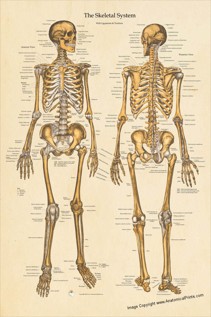
Human Skeletal Anatomy Poster Anterior and Posterior views ... from i.etsystatic.com The meniscus is a shock absorber that sits between the end of the thigh bone and the top of the shin bone. Jun 28, 2021 · 3 thenar muscles: See full list on kenhub.com Bones in human body is the solid structure that helps in making the physical appearance of the body. The muscle acts as the effort force; Understanding normal function of the knee joint can help you to address some of these common conditions. The synovial cells produce a slippery, viscous fluid called synovial fluid within the joint. Muscles propel the knee joint back and forth.
The patellar tendon on the front of the knee is part of the quadriceps mechanism.
Other smaller muscles and tendons surround the knee joint as well. The synovial cells produce a slippery, viscous fluid called synovial fluid within the joint. And the object being moved acts as the load. There are around 650 skeletal muscles within the typical human body. The quadriceps muscles provide strength and power with knee extension (straightening) and the hamstrings muscles allow for strength and power in flexion (bending). The tibia, femur, and patella, all are covered with a smooth layer of cartilage (see below) where they contact each other at the knee joint. A bursa is a structure in your body it is placed between two moving parts. Human anatomy diagrams show internal organs, cells, systems, conditions, symptoms and sickness information and/or tips for healthy living. The shin bone (tibia), the thigh bone (femur), and the kneecap (patella) are each important parts of the knee joint. Biology supplies, medical simulators, anatomical models, physics See full list on verywellhealth.com More images for human anatomy muscles and bones » The bone that the muscle moves acts as the lever;
There are two types of cartilage of the knee joint human muscles and bones. Cartilage is a resilient structure that resists damage, but when injured it has a difficult time healing.